- Imagej software cell staining manuals#
- Imagej software cell staining full#
- Imagej software cell staining series#
- Imagej software cell staining download#
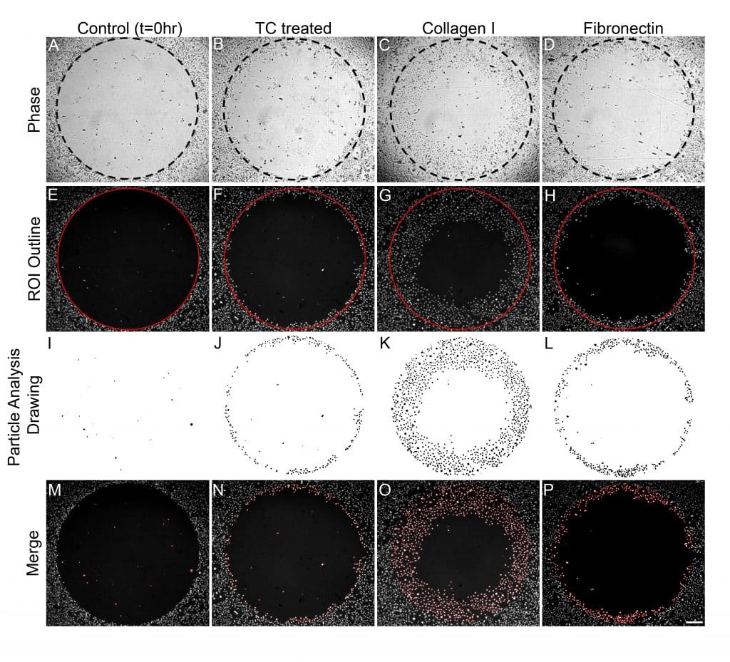
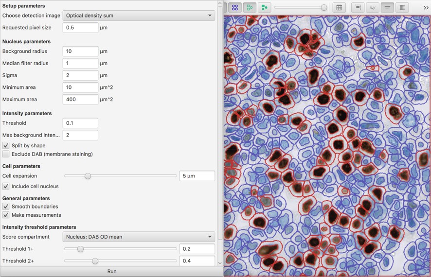
Existing image analysis programs, for example, CELLPROFILER and IMAGEJ, require pipeline/macro/plugin files that are specific to the cell/assay types, which is not available yet for the Transwell assays.

Such images may contain hundreds of cells and manually counting the number of them is not a trivial work. The number of cells that pass through the filter membrane from Boyden chamber is usually counted manually from the inverted microscopic images. Transwell Boyden chamber based cell migration/invasion assay is a simple and extensively used approach for the quantitation of cell motility in vitro. Cell migration/invasion is central to many physiological and pathological processes such as embryonic development, wound repair, and tumor metastasis. Cell invasion is similar to cell migration, except that it requires the cell to migrate through an extracellular matrix or basement membrane barrier by enzymatically degrading the barrier. IntroductionĬell migration is the movement of cells from one location to another generally in response to and toward specific external chemical signals. CELLCOUNTER will be helpful in streamlining the experimental process and improving the reliability of the data acquisition. The counting can be performed in batch, and the counting results can be visualized and further curated manually. We have therefore developed CELLCOUNTER, an application that is capable of recognizing and counting the total number of cells through an intuitive graphical user interface. The counting is usually performed manually, which is laborious and error prone. Cell motility is quantified by counting the number of cells that pass through the filter membrane.
Imagej software cell staining series#
if you want to break down the multidimension series e.g.Transwell Boyden chamber based migration/invasion assay is a simple and extensively used approach for the characterization of cell motility in vitro.convert the opened stack to hyperstack, do Image > Hyperstack > Stack to hyperstack., use the default xyczt order and fill in the appropriate c, z, t value.need to use View stack with: Standard ImageJ and Stack order: Default (xyzct).sometimes simultaneous multichannel files may open with channels interleaved, and here is a workaround.


click OK and wait for loci_tools' Bio-Formats Series Options dialog box which allows selection of specific contents.under Color options, check Autoscale if you want to use the min and max intensity values stored in the image.for Stack Viewing, set View stack with: Hyperstack.after a few moments of thinking, loci_tools' Bio-Formats Import Options dialog box will open.drag and drop the lif file onto ImageJ's status bar to open the file.some software cannot deal with 16-bit files, you can convert the image to 8-bit by doing Image > Type > 8-bit.click on Set and enter the desirable displayed values or click on Auto to use the min and max intensity values stored in the image.

Imagej software cell staining full#
*16-bit files with 12-bit resolution will appear dark unless the intensity is scaledĮ.g., set Minimum displayed value to 0 and Maximum displayed value to 4095 for full scale display the system outputs 12-bit data and images can be acquired with 8, 12 or 16-bit resolution.
Imagej software cell staining download#
Imagej software cell staining manuals#
There are excellent manuals and tutorials available on the NIH ImageJ website. Using ImageJ with file formats created on our microscopes


 0 kommentar(er)
